上海金畔生物科技有限公司代理New England Biolabs(NEB)酶试剂全线产品,欢迎访问官网了解更多产品信息和订购。
产品信息
5-methylcytosine (5-mC) is the predominant epigenetic mark in mammalian genomic DNA. 5-hydroxymethylcytosine (5-hmC) is a newly discoveredepigenetic modification that is presumably generated by oxidation of 5-mC by the TET family of cytosine oxygenases.1,2
Techniques exist that can identify 5-mC in genomic DNA, but the most commonly used method, bisulfite sequencing, is laborious and cannot distinguish between 5-mC from 5-hmC.3
The kit distinguishes 5-mC from 5-hmC by the addition of glucose to the hydroxyl group of 5-hmC via an enzymatic reaction utilizing T4 β-glucosyltransferase (T4-BGT). When 5-hmC occurs in the context of CCGG, this modification converts a cleavable MspI site to a noncleavable one.
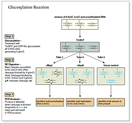
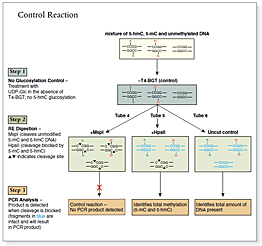
An overview of the detection procedure is summarized in Figure 1.
Control DNA Sequence 5´-CAGTGAAGTTGGCAGACTGAGCCAGGTCCCACAGATGCAGTGACCGGAGT CATTGCCAAACTCTGCAGGAGAGCAAGGGCTGTCTATAGGTGGCAAGTCA-3´
Control DNA substrates are synthetic 100 bp double stranded fragments containing a single MspI/HpaII site (CCGG). The three fragments are identical except for modification of the internal C in this site.
FW Primer Sequence 5´- CA GTG AAG TTG GCA GAC TGA GC -3´
REV Primer Sequence 5´- CTG ACT TGC CAC CTA TAG ACA GC -3´
产品类别:
Discontinued (<3 years)
特性和用法
需要但不提供的材料
Heat block or water bath (suitable for temperatures of 37°C, 40°C and 95°C) PCR materials:
Locus-specific primers, flanking a CCGG site of interest
0.2 ml strip tubes and caps for PCR 1.5 ml reaction tubes Molecular biology grade water
方法概述
方法概述
Step I: DNA Glucosylation Reaction with T4 β-glucosyltransferase (T4-BGT) Genomic DNA of interest is treated with T4-BGT, adding a glucose moeity to 5-hydroxymethylcytosine. This reaction is sequence-independent – therefore all 5-hmC will be glucosylated, unmodified or 5-mC containing DNA will not be affected.
Step II: Restriction Endonuclease DigestionMspI and HpaII recognize the same sequence (CCGG) but are sensitive to different methylation status. HpaII cleaves only a completely unmodified site: any modification (5-mC, 5-hmC or 5-ghmC) at either cytosine blocks cleavage. MspI will recognize and cleave 5-mC and 5-hmC, but not 5-ghmC.
Step III: Interrogation of the Locus by PCR as little as 20 ng of input DNA can be used. Amplification of the experimental (glucosylated and digested) and control (mock glucosylated and digested) target DNA with primers flanking a CCGG site of interest (100–200 bp) is performed. If the CpG site contains 5-hydroxymethylcytosine, a band is detected after glucosylation and digestion, but not in the non-glucosylated control reaction (see Figure 2). Real time PCR will give an approximation of how much hydroxymethylcytosine is in this particular site.
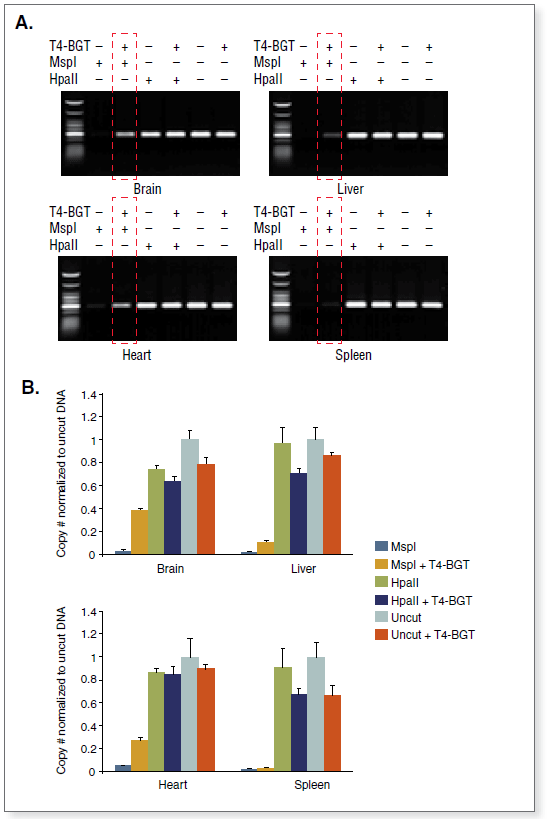
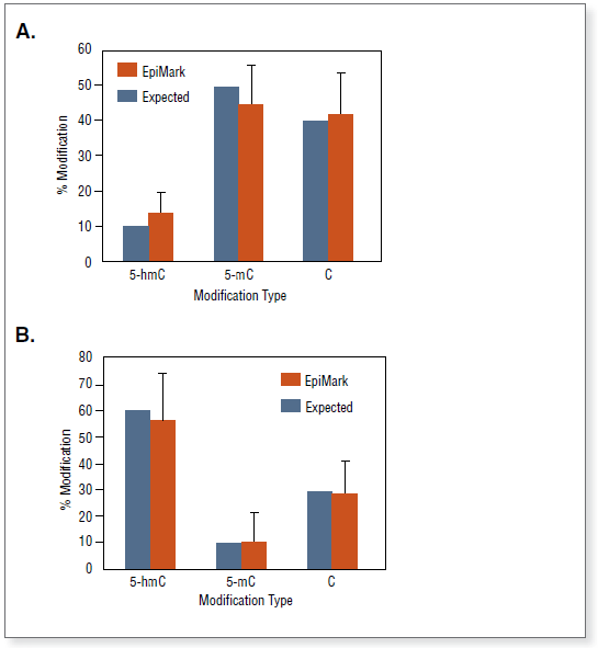
Figure 1a: Experimental Overview The DNA of interest is treated with T4 β-Glucosyltransferase (T4-BGT) and UDP-Glucose (UDP-Glc). T4-BGT transfers glucose from UDP-Glc onto 5-hydroxymethylcytosine (generating glucosylated 5-hydroxymethylcytosine [5-ghmC]). MspI cuts DNA containing 5-hmC, but does not cut 5-ghmC containing sites; in contrast, HpaII is blocked by any of these modifications. Presence of 5-hmC and 5-mC can be determined by PCR analysis.Figure 1b: Experimental OverviewThe DNA of interest is digested following a control reaction with UDP-Glucose (UDP-Glc) and no T4 β-Glucosyltransferase (T4-BGT), leaving 5-hmC unmodified. MspI cleaves unmodified, 5-mC and 5-hmC DNA, while HpaII cleaves only unmodified DNA.Figure 2: Comparison of 5-hydroxymethylcytosine amounts at locus 12 in different mouse Balb/C tissue samples. (A) End-point PCR. (B) Real time PCR.DNA from four mouse tissues was analyzed. For comparative purposes, real time PCR data were normalized to uncut DNA. A standard curve was used to determine copy number. The samples could be normalized by dividing the copy number of samples No 1-6 by the copy number of the control that is undigested (No 5). Boxed gel lane shows variation in 5-hmC present.Figure 3: High sensitivity 5-hydroxymethylcytosine detection achieved by the EpiMark kit.100 bp unmodified, 5-mC, and 5-hmC control DNAs were mixed in different ratios (blue bars), and then measured with the EpiMark hydroxymethylated DNA detection kit (orange bars). Error bars represent the standard deviation of four independent experiments.